Daniel Konicek Contributing Writer
Understanding the organs in the breast is an important first step.
A typical breast consists of three 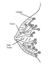 things, the lobules, ducts, and stroma. A lobule is a small gland, basically a tiny factory, responsible for producing milk, and they are spread throughout the breast. Connecting the lobules are ducts, which are tiny tubes that carry the milk from the glands to the nipple.
Cancer cells will typically begin to grow into a tumor within the lining of the ducts and lobules of the breast. The rest of the breast consists of stroma, essentially the fatty tissue containing blood vessels and connective tissue that surrounds the lobules and ducts.
When cancer begins, it is usually a small tumor in the lining of an organ, called a carcinoma. Until the cells spread outside the tumor, it is called pre-invasive, or carcinoma in situ, and is the optimal time for operation.
Finding these small lumps is the primary purpose of mammograms, MRIs and clinical breast exams. If the cancer can be treated before it spreads and becomes invasive, the chances of survival are drastically increased.
Unfortunately, it is very easy for breast cancer cells to begin spreading. There are a large amount of lymph nodes present in or near the breast, responsible for sending white blood cells, hormones and waste products through the body. If the cancer spreads to these nodes, it can quickly travel and grow in places like the brain, bone marrow, lung or liver.
Detecting carcinomas, or rarer cancers growing in the stroma, before they become invasive is absolutely essential to the health of woman.
The American Cancer Society recommends regular mammograms, self exams and clinical breast exams for this very reason. Each type of test has different strengths and weaknesses, so a well-rounded and consistent approach is a necessity. Many factors, like breast density and family history, can change how often exams are recommended.